
What is a microscope? In the rigorous pursuit of scientific truth, this instrument is far more than a mere magnifying tool; it is a sophisticated optical sentinel that bridges the chasm between human sensory limitations and the fundamental architecture of matter. Since the early 17th century, when pioneers like Antonie van Leeuwenhoek first interrogated drops of water to reveal a “vast universe” of microorganisms, the microscope has served as the primary vehicle for human progress in biology, medicine, and materials science. It is the definitive apparatus that allows us to transition from speculative observation to empirical data collection at the cellular and crystalline levels.
Defining the Instrument: What is a Light Microscope?
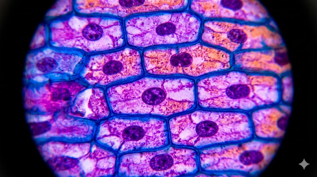
To understand the core of modern microscopy, we must first address the most prevalent form: what is a light microscope? Technically referred to as a compound optical microscope, this device utilizes visible light and a series of high-precision glass lenses to resolve images of small objects. The fundamental physics governing its operation rely on the refraction of light—the bending of light waves as they pass from one medium (air) through another (glass) with a different refractive index.
A light microscope typically operates by passing light through a thin, translucent specimen. This light is then gathered by the objective lens, which creates a magnified “real image” within the body tube. This image is further magnified by the ocular lens (eyepiece), resulting in the final “virtual image” perceived by the observer’s eye or a digital sensor. The true power of a light microscope is measured not just by its magnification—the ratio of image size to object size—but by its resolution. Resolution is the ability of the optical system to distinguish two separate points as distinct entities, a limit dictated by the wavelength of light and the numerical aperture of the lenses.
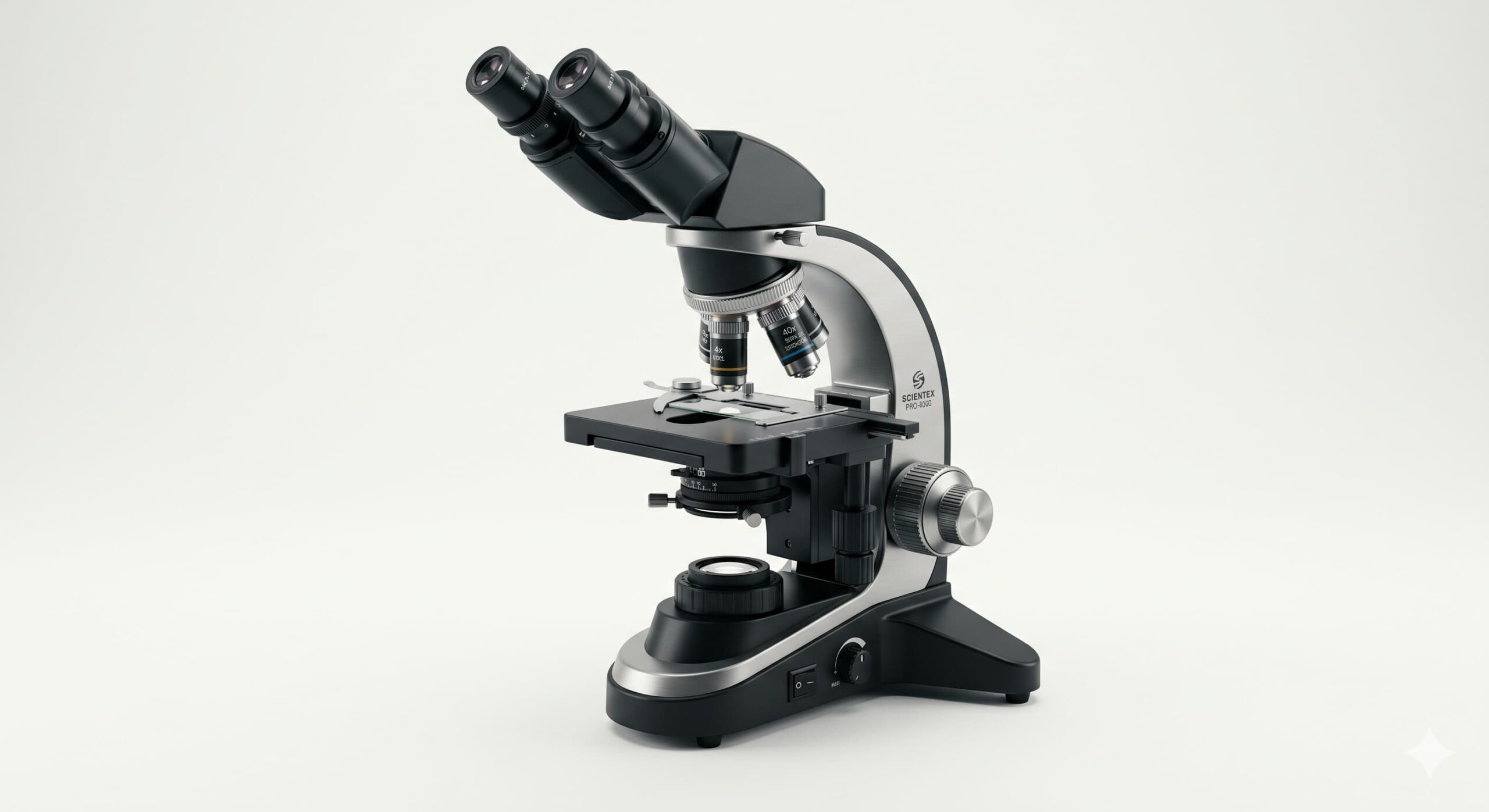
The Anatomy of Precision: Parts of a Microscope
A scientific instrument is only as reliable as its mechanical and optical components. The parts of a microscope are divided into two primary categories: the mechanical framework and the optical train. Understanding these components is essential for any researcher aiming to achieve high-quality micrographs.
The Mechanical Framework
- The Base and Arm: These provide the structural integrity and stability required to prevent vibrations, which can be catastrophic at high magnifications.
- The Stage: The horizontal platform where the specimen is placed. In professional research models, this is a mechanical stage equipped with X-Y translational controls for precise movement.
- Adjustment Knobs: The coarse and fine adjustment knobs move the stage (or the body tube) vertically to bring the specimen into focus. The fine adjustment is critical when working with high-power objectives where the depth of field is measured in micrometers.
The Optical Train
- The Illuminator: The light source, typically a high-intensity LED or halogen bulb located in the base.
- The Condenser: A lens system located beneath the stage that concentrates light into a focused beam on the specimen.
- The Iris Diaphragm: Located within the condenser, it controls the contrast and the angle of the light cone, a vital step in optimizing image quality.
- The Objectives: These are the most critical components. Most microscopes feature a revolving nosepiece carrying three to five objective lenses of varying powers (typically 4x, 10x, 40x, and 100x).
- The Eyepiece (Ocular): The lens the user looks through, usually providing an additional 10x magnification.
Functional Utility: What is a Microscope Used For?
The question of what is a microscope used for spans nearly every branch of modern science. Its utility is not confined to a single discipline but serves as a universal tool for morphological and structural analysis.
- Clinical Pathology and Medicine: In the medical field, microscopy is the gold standard for diagnosing diseases. From identifying malignant cells in a tissue biopsy to detecting parasitic infections in blood smears, the microscope provides the empirical evidence required for life-saving interventions.
- Materials Science and Metallography: Engineers utilize microscopy to examine the grain structures of metals and the integrity of polymers. By observing the microstructure, scientists can predict the mechanical properties of an alloy, such as its hardness, ductility, and susceptibility to fatigue.
- Forensic Investigation: Trace evidence—such as hair fibers, soil particles, or gunshot residue—is analyzed microscopically to link suspects to crime scenes with scientific certainty.
- Environmental Science: Microbiologists monitor water quality by identifying protozoa and algae populations, which serve as bio-indicators of ecosystem health.
Operational Methodology: How Do You Switch Objectives on a Microscope?
Achieving optimal visualization requires a systematic approach to magnification. A common point of inquiry for novice researchers is how do you switch objectives on a microscope? The procedure is governed by the use of the revolving nosepiece.
To transition from a lower magnification to a higher one, the operator must grasp the knurled outer ring of the revolving nosepiece and rotate it until the desired objective “clicks” into the optical path. It is a critical professional standard to never pull on the objective tubes themselves, as this can misalign the delicate internal optics.
Most modern research microscopes are “parfocal,” meaning that once the specimen is in focus at a lower power, it should remain largely in focus when switching to a higher power, requiring only a slight adjustment with the fine focus knob. This systematic escalation—starting at 4x to locate the area of interest before moving to 40x or 100x for detailed analysis—is the hallmark of a disciplined microscopist.
Beyond the Visible: The Evolution of Microscopy
While the light microscope is the workhorse of the laboratory, the quest for higher resolution led to the development of electron microscopy. Because electrons have a much shorter wavelength than visible light, instruments like the Scanning Electron Microscope (SEM) and the Transmission Electron Microscope (TEM) can resolve structures at the nanometer and even atomic scales.
These advancements allow us to interrogate the surface of a virus, the lattice structure of a semiconductor, or the internal organelles of a cell in three dimensions. However, the light microscope remains irreplaceable due to its ability to observe living specimens in real-time, its ease of sample preparation, and its cost-effectiveness.
Conclusion: The Unending Quest for Clarity
In summary, when we ask what is a microscope, we are describing the ultimate instrument of human curiosity. It is a fusion of physics, engineering, and art that allows us to peer into the “hidden” world that dictates the laws of our macroscopic reality. Whether it is used for identifying a new bacterial strain or ensuring the safety of a bridge’s steel girders, the microscope remains the most vital tool in the scientist’s arsenal. As technology advances into the realms of super-resolution and automated imaging, the microscope will continue to evolve, forever pushing the boundaries of what is visible and what is known.


Leave a Reply