
The microscope camera is the indispensable bridge between optical phenomena and digital quantification. In the modern laboratory, the act of “seeing” has transitioned from a subjective, fleeting observation through an eyepiece to an objective, permanent data record. As we move through 2026, the integration of high-resolution sensors with precision optics has fundamentally altered the workflow of researchers across disciplines—from clinical pathology to advanced materials science. For the contemporary scientist, the choice of a digital acquisition system is as critical as the selection of the objective lenses themselves.
From Eyepiece to Sensor: The Evolution of Visualization
Historically, microscopy was a solitary endeavor. Scientists would peer through oculars and manually sketch their findings. The introduction of film photography brought a degree of permanence, but it was the advent of the digital camera microscope ecosystem that truly democratized scientific visualization. Today, the ability to capture, share, and analyze images in real-time has accelerated the pace of global collaboration.
At its core, a microscope camera is a specialized imaging device designed to interface with the optical path of a microscope to capture the “real image” formed by the objective and tube lenses. Unlike consumer photography, where artistic flair is often prioritized, microscopy imaging is governed by the laws of Nyquist sampling, signal-to-noise ratios (SNR), and quantum efficiency. The goal is not merely to create an image, but to produce a mathematically accurate representation of the specimen.
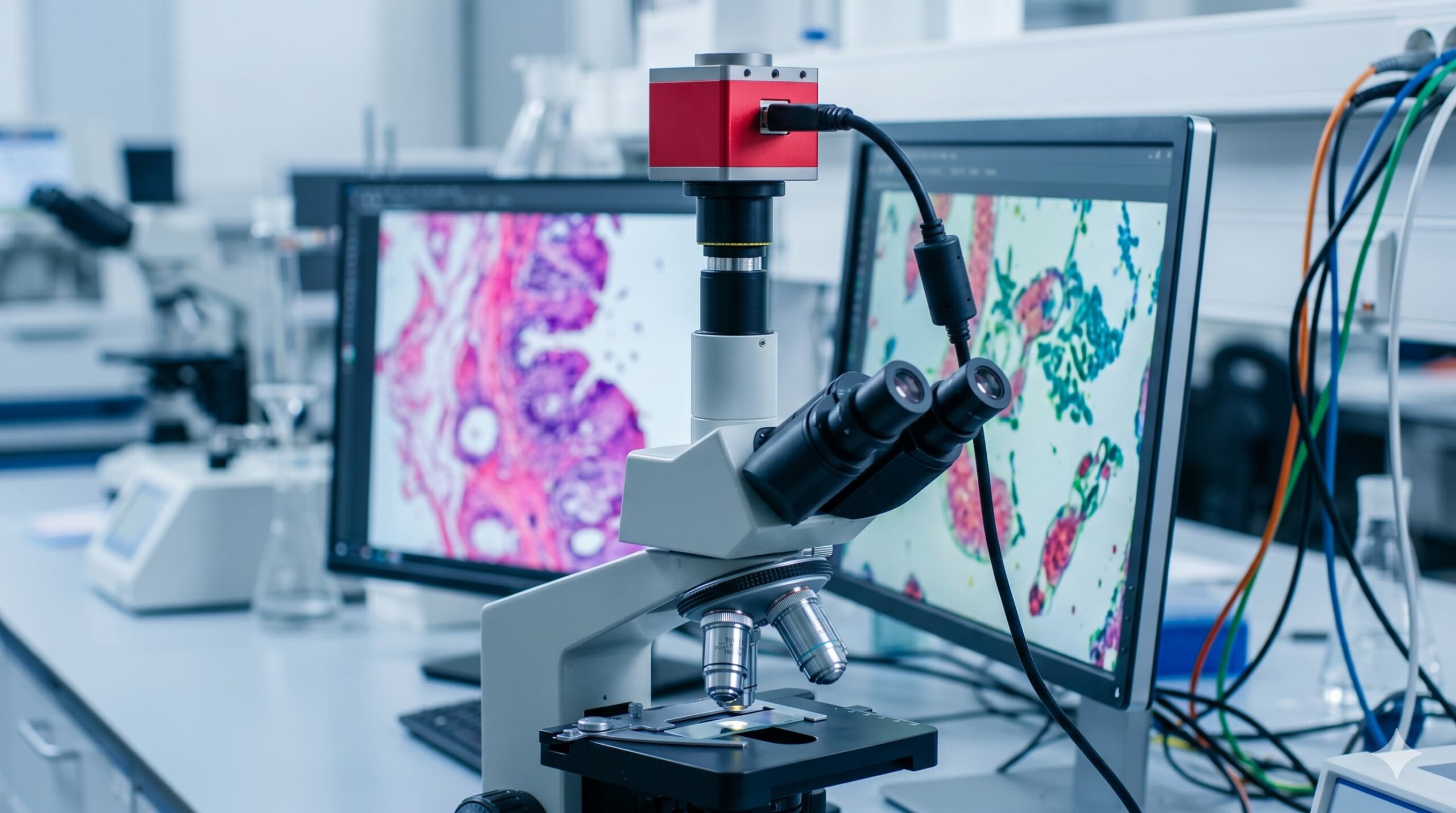
The Hardware Interface: The Trinocular Microscope
For a professional-grade imaging setup, the trinocular microscope is the industry standard. While binocular models are sufficient for visual inspection, they require the removal of an eyepiece to accommodate a camera, which disrupts the workflow. A trinocular head features a third vertical port—a dedicated optical path for the sensor.
The integration usually involves a “C-mount” or “F-mount” adapter. These adapters often contain a reduction lens (e.g., 0.5x or 0.75x) to match the sensor’s size with the microscope’s field of view. A well-aligned trinocular microscope ensures “parfocality,” meaning that when the image is in focus for the observer’s eyes, it is simultaneously in focus for the camera sensor. This synchronization is vital for high-throughput environments where time-lapse imaging or rapid documentation is required.
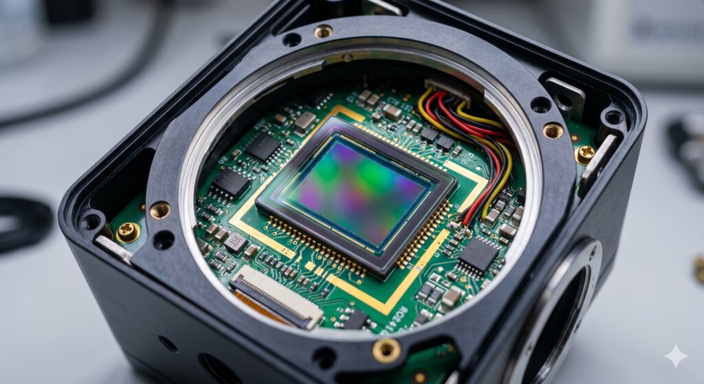
Sensor Technology: CMOS vs. CCD
The heart of every microscope camera is its sensor. For decades, Charge-Coupled Devices (CCD) were the gold standard due to their high sensitivity and low noise. However, the paradigm has shifted. Modern Scientific CMOS (sCMOS) sensors now offer comparable—and often superior—performance with significantly higher frame rates and wider dynamic ranges.
In a scientific context, pixel size is a critical variable. While a microscope for kids might prioritize a high “megapixel” count as a marketing gimmick, professional cameras focus on pixel quality. Larger pixels (e.g., 6.5 microns) can collect more photons, providing a higher SNR, which is essential for low-light applications like fluorescence microscopy. Smaller pixels, conversely, provide higher spatial resolution, provided they do not exceed the resolving power of the microscope’s objective.
The Gold Standard: The Nikon Microscope Camera
When discussing high-fidelity imaging, the nikon microscope camera series often serves as a benchmark for the industry. These systems are engineered to integrate seamlessly with the “CFI60” infinity-corrected optical system. The precision of a nikon microscope camera lies in its ability to maintain color fidelity and geometric accuracy across the entire sensor area.
Professional systems from top-tier manufacturers focus heavily on bit depth. While a standard JPEG image uses 8 bits per channel (256 levels of gray), a scientific camera often captures data at 12 or 16 bits (over 65,000 levels). This “depth” allows researchers to distinguish between subtle variations in stain intensity or material density that would be invisible to the human eye or a consumer-grade sensor.
Educational vs. Professional: The Microscope for Kids
The technology has scaled so efficiently that even the microscope for kids market has seen a digital revolution. In an educational setting, the primary goal of a microscope camera is engagement. These entry-level devices are often all-in-one units where the sensor is integrated into the head, allowing students to view live “pond life” or plant cells on a tablet or laptop.
While the sensor in a microscope for kids lacks the cooled housing (to reduce thermal noise) or the high quantum efficiency of a research-grade sCMOS, it fulfills a vital role: training the next generation of scientists in digital literacy. It introduces the concepts of focus, exposure, and digital scaling, which are the foundations of professional microscopy.
Image Acquisition and Data Integrity
A scientist does not simply “take a photo.” The process of digital acquisition involves several critical steps to ensure data integrity:
- Calibration: Every microscope camera must be calibrated using a stage micrometer. This links pixel dimensions to physical units (microns), allowing for accurate measurements of grain size, cell diameter, or crack length.
- White Balance and Background Subtraction: To ensure color accuracy, the camera must be balanced against the light source. Background subtraction (or “Flat Field Correction”) removes uneven illumination or dust shadows from the final data.
- Frame Rate and Shutter Type: For live specimens or moving parts, a “Global Shutter” is preferred over a “Rolling Shutter” to avoid the spatial distortion of moving objects.
The Role of Software in Digital Microscopy
While the hardware captures the photons, the software transforms those signals into knowledge. Modern digital camera microscope suites facilitate advanced techniques such as:
- Extended Depth of Field (EDF): Combining multiple images at different focal planes to create a single sharp image of a 3D object.
- Image Stitching (Mosaicing): Merging hundreds of individual fields of view into a single gigapixel “map” of a large specimen.
- Automated Quantification: Using algorithms to count thousands of cells or particles in seconds, a task that would take a human researcher days to complete.
Conclusion: The Precision of the Digital Eye
In summary, the microscope camera is far more than an accessory; it is a precision instrument that defines the capabilities of the modern laboratory. From the simple CMOS sensors in a microscope for kids to the cooled, high-speed arrays in a nikon microscope camera, these devices have transformed microscopy into a quantitative science.
By understanding the synergy between the trinocular microscope and the digital camera microscope interface, scientists can unlock insights into the hidden architecture of the world. As we look toward the future, the integration of Artificial Intelligence with these imaging systems promises to further push the boundaries of what can be seen, measured, and understood.


Leave a Reply