
Anyone who has ever looked through a high-powered microscope or attempted extreme macro photography has encountered the exact same frustrating barrier: the agonizingly thin slice of focus. You perfectly adjust your lens to see the peak of a microscopic structure, only to realize that the base and the background have dissolved into an unrecognizable blur.
This optical limitation is governed by the laws of physics, but thanks to modern computational photography and advanced software algorithms, we are no longer bound by it. The solution is a revolutionary technique known as Extended Depth of Field (EDOF).
Whether you are working in metallography analyzing fractured steel parts, examining complex printed circuit boards (PCBs), or documenting biological specimens, understanding and utilizing EDOF is crucial for modern digital imaging. In this comprehensive guide, we will explore exactly what EDOF is, the physics behind why we need it, and how advanced software transforms blurry captures into perfectly sharp, information-rich images.
The Physics of the Problem: Why Does Focus Disappear?
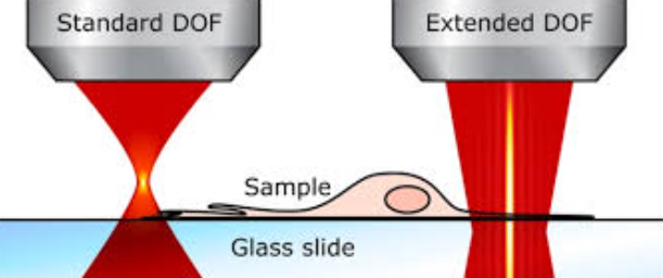
To appreciate the magic of EDOF, we first need to understand the concept of standard Depth of Field (DoF). In optics, the depth of field is the distance between the nearest and the furthest objects that are in acceptably sharp focus in an image.
When you use a standard camera with a wide-angle lens, your DoF might be several meters or even miles deep. However, as you increase your magnification, the depth of field shrinks exponentially. In the realm of optical microscopy and macro photography, depth of field is often measured not in inches, but in micrometers ($\mu m$).
This drastic reduction is primarily tied to the Numerical Aperture (NA) of the objective lens. To achieve high resolution and gather enough light at high magnifications, lenses require a high NA. Unfortunately, physics dictates a cruel trade-off: as Numerical Aperture increases, the Depth of Field decreases.
If you are inspecting a three-dimensional object—such as a rough metallic fracture in metallography or a spherical biological cell—a high-magnification lens can only focus on a tiny horizontal slice (the Z-plane) of that object at any given time. The rest of the topography remains blurred, obscuring vital data and making comprehensive analysis impossible.
What is Extended Depth of Field (EDOF)?
Extended Depth of Field (EDOF), frequently referred to in the photography world as focus stacking or Z-stacking, is a digital image processing technique that combines multiple images taken at different focal distances to generate a single, fully in-focus composite image.
Instead of trying to engineer a physical lens that defies the laws of physics (which is impossible), EDOF relies on computational power. By capturing a sequence of images—moving the focus slightly from the highest point of the sample down to the lowest point—you collect all the necessary sharp data across various Z-axis planes. Advanced algorithms then analyze this “stack” of images, extract the sharpest pixels from each individual frame, and seamlessly stitch them together.
The result is a final two-dimensional image that boasts a depth of field that would be physically impossible to achieve with a single optical exposure.
How the EDOF Software Works: Behind the Algorithms
The process of generating an EDOF image might seem instantaneous to the end-user, especially when using premium imaging suites (such as Microvision or modern metallography software), but the underlying mathematical operations are incredibly complex. The workflow generally consists of four critical stages:
1. Image Acquisition (Building the Z-Stack)
The first step is capturing the raw data. The camera takes a picture, the lens (or the motorized microscope stage) moves slightly along the Z-axis, and another picture is taken. This process repeats until the entire vertical topography of the subject has been photographed. Depending on the depth of the sample and the magnification, a stack can consist of anywhere from 5 to over 500 individual images.
2. Image Alignment and Registration
When a lens changes focus, it often creates a subtle optical effect known as “focus breathing”—the magnification changes very slightly, making objects appear to grow or shrink. If the software simply stacked the images as-is, the final result would be misaligned and distorted. EDOF algorithms must first meticulously align and scale every image in the stack so that the underlying structures match perfectly from frame to frame.
3. Contrast Evaluation and Pixel Selection
This is the core of the EDOF algorithm. How does a computer know what is “in focus” and what is “blurry”? It looks for local contrast. Blurry areas feature smooth, gradual transitions between pixels. Sharp, in-focus areas feature abrupt, high-contrast changes between adjacent pixels (sharp edges). The software analyzes the entire stack pixel-by-pixel (or block-by-block), identifying the specific layer where each pixel exhibits the highest local contrast.
4. Blending and Artifact Removal
Once the sharpest pixels are identified, the software blends them together into a final map. High-end EDOF software includes advanced smoothing algorithms to ensure that the transitions between different focal layers are invisible, eliminating “halos” or weird artifacts that can sometimes occur around overlapping edges.
Real-World Applications: Where EDOF is Indispensable
The ability to generate completely focused images of microscopic 3D objects has revolutionized numerous industries.
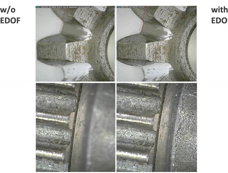
- Metallography and Material Science: When conducting precise metallography analysis, examining metal fatigue, corrosion, or fractured surfaces often reveals a highly uneven sample topography. Standard microscopy provides a frustratingly narrow view. EDOF allows engineers to view a highly detailed, deeply focused image of the entire fracture, making it drastically easier to determine the cause of material failure and ensure compliance with strict ASTM standards.
- Electronics and PCB Inspection: Modern microchips and printed circuit boards feature multi-layered topographies. Solder joints, microscopic wires, and silicon components exist on different physical planes. EDOF is essential for quality control in electronics manufacturing, allowing inspectors to view entire component assemblies in sharp focus simultaneously.
- Entomology and Life Sciences: Biological samples are rarely perfectly flat. Trying to photograph an insect’s head under a microscope usually results in the eyes being in focus while the antennae blur away. EDOF allows biologists to create stunning, fully focused visualizations of complex anatomical structures for research and publication.
Hardware vs. Software: Choosing the Right Setup
Achieving excellent Extended Depth of Field requires a synergy between hardware and software.
For occasional use, you can manually turn the fine-focus knob on a microscope while snapping photos, and then feed those images into standalone focus-stacking software. However, manual turning is prone to human error and uneven spacing between focal planes.
For professional, industrial, or high-volume environments, motorized Z-axis drives are highly recommended. These automated systems communicate directly with the microscopic imaging software. The user simply defines the top focal point and the bottom focal point, and the software automatically calculates the optimal step size, drives the motor, captures the images, and renders the final EDOF image in seconds.
Conclusion
Extended Depth of Field (EDOF) is much more than just a neat software trick; it is an essential analytical tool that has profoundly changed how we interpret the microscopic world. By effectively neutralizing the restrictive physical limitations of optical lenses, EDOF provides scientists, engineers, and photographers with clear, comprehensive, and accurate visual data. As computational imaging continues to evolve, the algorithms behind EDOF will only become faster and more precise, pushing the boundaries of what we can discover under the lens.
Leave a Reply