
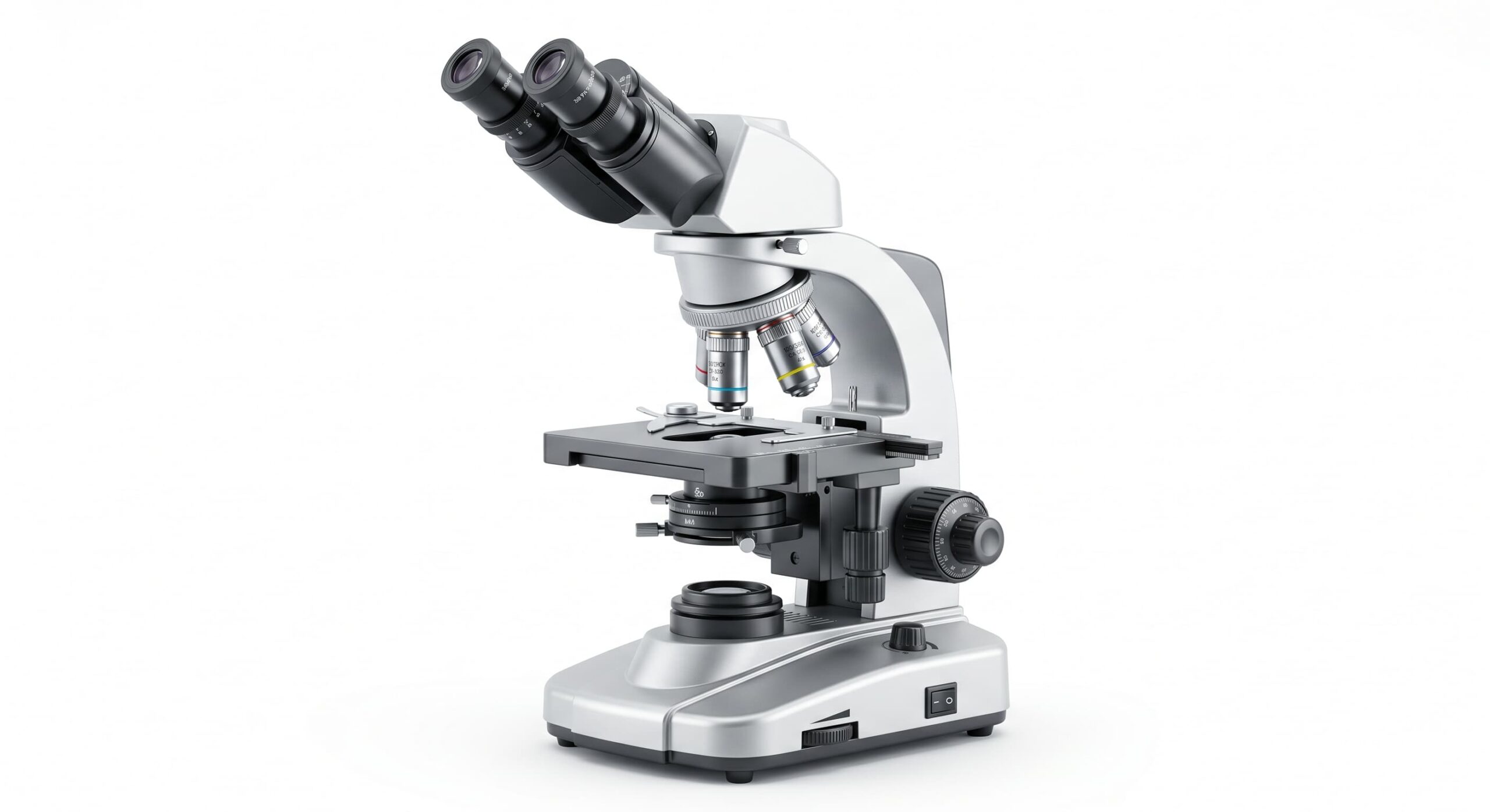
The bright field microscope remains the foundational instrument of the modern laboratory, acting as the primary vehicle for histological, microbiological, and cytological discovery. Within the rigorous framework of materials science and biological research, the ability to visualize the internal and external architecture of a specimen is predicated on the mastery of light transillumination. While the 21st century has introduced a myriad of specialized modalities—such as fluorescence, phase-contrast, and confocal microscopy—the bright field microscope continues to be the workhorse of clinical diagnostics and academic research due to its relative simplicity, cost-effectiveness, and the high-fidelity images it provides when properly calibrated.
The Physics of Transillumination: How the Bright Field Microscope Functions
At its most fundamental level, the bright field microscope operates on the principle of light transmission. In this configuration, a light source (typically a high-intensity LED or halogen bulb) is positioned beneath the specimen. The light passes through a series of lenses—starting with the condenser—before traversing the specimen. The “bright field” designation refers to the appearance of the resulting image: the specimen appears dark or colored against a brilliantly illuminated background.
The quality of the image produced by a digital bright field microscope is not merely a product of the objective lens’s magnification power. It is a complex interplay of illumination, contrast, and resolution. When light waves encounter a specimen, they are absorbed, reflected, or refracted. The differences in these interactions across various regions of the specimen create the contrast that allows the human eye or a digital sensor to distinguish one structure from another.
The Quest for Resolution: Abbe’s Law and Numerical Aperture
In professional microscopy, magnification is often a secondary concern compared to resolution. Resolution is defined as the minimum distance between two points at which they can still be distinguished as separate entities. This limit was scientifically quantified by Ernst Abbe in 1873. According to Abbe’s Law, the resolution of a bright field microscope is limited by the wavelength of light and the numerical aperture (NA) of the objective and condenser lenses.
The Numerical Aperture is a dimensionless number that characterizes the range of angles over which the system can accept or emit light. To maximize resolution, researchers often utilize immersion oil when working at high magnifications (typically 100x). Immersion oil has a refractive index similar to glass (approximately 1.51), which prevents the light from refracting as it exits the coverslip and enters the air. By bridging this gap with oil, the NA is significantly increased, allowing the bright field microscope to reach its theoretical resolution limit of approximately 0.2 micrometers.
Mastering Contrast: The Role of Chemical Staining
The primary limitation of the bright field microscope is the inherent transparency of biological specimens. Most living cells consist largely of water and offer very little refractive difference from their surrounding medium. To overcome this, scientists employ chemical staining techniques.
Staining serves to increase the contrast of specific cellular or extracellular components. For example:
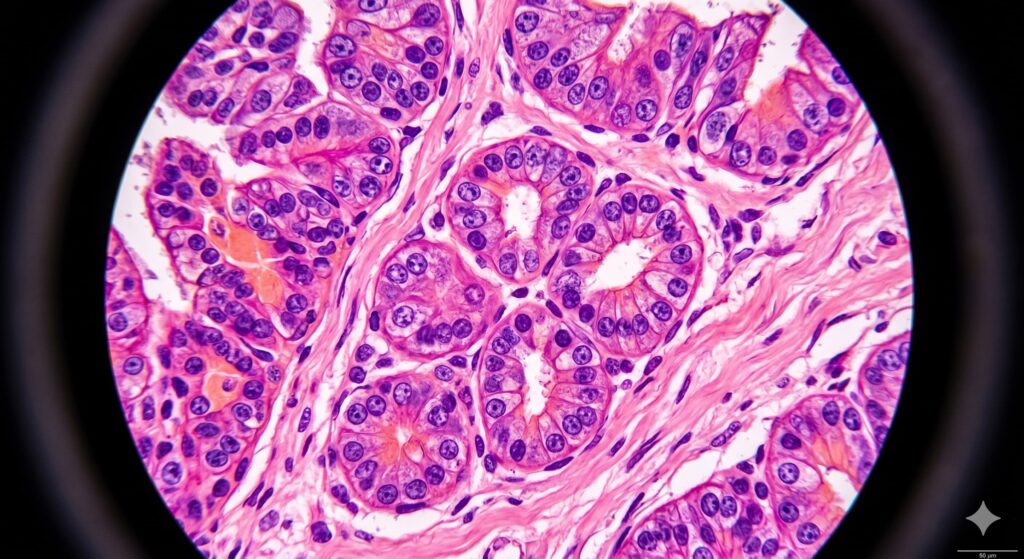
- Hematoxylin and Eosin (H&E): The gold standard in pathology. Hematoxylin stains acidic structures (like the nucleus) deep blue, while Eosin stains basic structures (like the cytoplasm) pink.
- Gram Stain: Used in microbiology to differentiate bacteria based on the chemical and physical properties of their cell walls.
- Masson’s Trichrome: Used to distinguish between muscle fibers and connective tissue in histological sections.
In a digital bright field microscope system, the sensor must be calibrated to represent these colors with extreme fidelity, as the saturation and hue of a stain can provide critical diagnostic information regarding the health or pathology of a tissue sample.
The Precision of Koehler Illumination
A hallmark of professional scientific microscopy is the implementation of Koehler Illumination. Named after August Koehler, this technique ensures that the illumination of the specimen is perfectly even and free from the glare of the light source’s filament or LED architecture.
To achieve Koehler Illumination on a bright field microscope, the researcher must precisely align the field diaphragm and the condenser. This process centers the light path and focuses it directly into the plane of the specimen. Proper Koehler alignment eliminates diffraction artifacts and maximizes the effective numerical aperture of the system. Without this rigorous calibration, the resulting image—no matter the quality of the camera—will suffer from “vignetting” (dark corners) and a significant loss of fine detail.
The Digital Transition: CMOS Sensors and Quantitative Analysis
In the modern era, the digital bright field microscope has largely replaced the traditional ocular-only setup. By integrating high-resolution CMOS (Complementary Metal-Oxide-Semiconductor) sensors, the microscope transitions from a viewing tool to a data-gathering instrument.
A digital bright field microscope allows for several advanced scientific functions:
- Automated Morphometry: Measuring the area, perimeter, and diameter of thousands of cells or particles in seconds.
- Colorimetric Quantification: Using digital values to determine the intensity of a specific stain, which can indicate the concentration of a protein or mineral within the sample.
- Digital Stitching: Capturing hundreds of individual bright field images and merging them into a single, gigapixel “map” of a large tissue section or metallurgical sample.
For the materials scientist, the bright field microscope is often used in a “reflected” light configuration to examine the polished surfaces of metals (metallography). Here, the light hits the specimen from above and reflects back into the objective, revealing grain boundaries, inclusions, and phase distributions that are critical to understanding a material’s mechanical properties.
Maintenance and Calibration: The Scientist’s Responsibility
To maintain research-grade performance, the bright field microscope requires meticulous care. Dust on the internal lenses, oil residue on the 40x objective (which is usually a “dry” lens), and misalignment of the stage can all introduce errors into a scientific study.
Regular calibration using a certified stage micrometer is mandatory. This ensures that the digital pixels on the sensor correspond to real-world dimensions (microns). In a peer-reviewed environment, an image without a clear, calibrated scale bar is considered anecdotal rather than empirical. Furthermore, the light source should be checked for “color temperature” consistency, as shifts in the LED’s spectral output can alter the perceived results of a stained specimen over time.
Conclusion: The Enduring Legacy of Bright Field Optics
In conclusion, the bright field microscope is far from an archaic technology. It is a refined optical system that remains the first point of entry into the micro-world. Whether it is used to identify a malignant cell in a hospital or to verify the grain size of a new aerospace alloy, its role in the scientific method is irreplaceable.
By understanding the physics of light, the mathematics of numerical aperture, and the necessity of Koehler illumination, the scientist ensures that the digital bright field microscope provides a clear, unbiased window into the hidden structures of our reality. As imaging software and sensor technology continue to evolve, the clarity of the bright field will only become more vital to the advancement of human knowledge.


Leave a Reply